3D Printing of Multi-layer Scaffolds to mimic Patient Specific Implants:
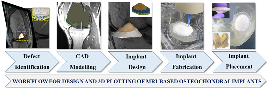
Customers at University Hospital of TU Dresden conducted a research study for the future therapy of osteochondral defects. They developed a workflow, based on a magnetic resonance imaging (MRI) dataset derived from a real clinical case. MRI provides more information on cartilage tissue and muscles, surrounding the defect area; whereas X-ray based CT provides a higher resolution but gives contrast for bone structures only.
The GeSiM BioScaffold printer BS3.1 was used to print a two-phase tissue scaffold (Step 4 of the workflow) with a design based on segmented MRI data of the lesioned zone. By extrusion-based printing, the 3D structure combined layers of an alg/MC (Alginate Methylcellulose) hydrogel for the cartilage zone with more rigid CPC (Calcium Phosphate Cement) to restore the subchondral bone portion. With this material combination whose suitability for bioprinting had been proven by the group before, even patient’s own cells can be directly embedded in the print process.
The final implant was tested by fitting it into a model of the actual defect geometry. For that purpose, an SLA model with the segmented data of the lesioned femoral condyle (FC) was created; it comprised particularly the area adjacent to the defect the implant was made for. The fitting process was supported by a case specific surgical plunger, made by SLA as well.

The GeSiM BioScaffold Printer BS3.1 was used for implant manufacturing (Step 4). The software of the instrument sliced the original STL data and defined parameters for the inner porosity.
The results show a perfect geometric fit of the implant and the defect model. The clinical performance of the generated implants still needs to be evaluated. However, the authors of the study are convinced, that in the future the demonstrated interdisciplinary process can be transferred to other patients and other clinical situations as well.
David Kilian et.al: 3D printing of patient-specific implants for osteochondral defects: workflow for an MRI-guided zonal design
Courtesy of: Centre for Translational Bone, Joint and Soft Tissue Research, Technische Universität Dresden

