Advancing Cancer Research with 3D Bioprinting Realistic Models:
Cancer progression is influenced by complex interactions between cancer cells and their surrounding stromal environment. Traditional 2D cell cultures and animal models have limitations in capturing this complexity. Therefore, researchers are increasingly adopting 3D in-vitro models that more accurately mimic the tumor microenvironment.
A team of researchers at McGill University has made significant strides in this area by utilizing 3D extrusion bioprinting with a composite bioink containing reinforced decellularized extracellular matrix (ECM) hydrogel to fabricate an in-vitro model of head and neck cancer. This model incorporates fibroblasts, representing the stromal component, and HNSCC cells, representing the tumor cells.
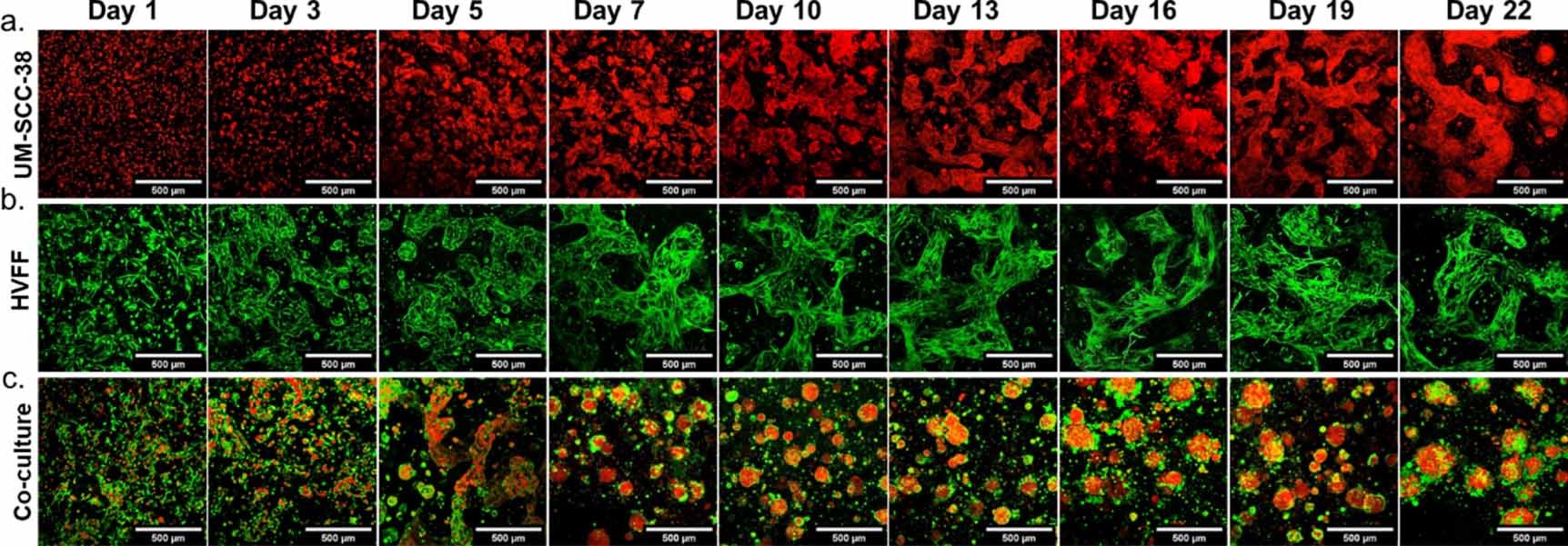
The GeSiM BioScaffolder played a crucial role in the 3D extrusion bioprinting process, ensuring precise cell placement and bioink deposition. Topographical characterization revealed that the bioink formed a fibrous network with nanometer-sized pores, closely mimicking the natural ECM. Over time, this bioink supported the development of multicellular spheroids, with cancer cells forming the core and fibroblasts surrounding them, effectively replicating the tumor-stroma interface.
Moreover, the model facilitated the quantification of matrix metalloproteinases (MMPs), specifically MMP-9 and MMP-10, which showed significant differences compared to control groups. This capability underscores the model’s potential for studying the dynamic interactions within the tumor microenvironment.
This 3D bioprinted model offers several key advantages for cancer research. It provides a realistic platform for investigating cancer-stromal interactions, enabling researchers to explore how stromal cells influence tumor behavior and vice versa. Additionally, the model allows for nondestructive longitudinal monitoring of changes over time, making it ideal for testing drug responses within a more realistic microenvironment.
The development of this 3D bioprinted model marks a significant advancement in cancer research. By providing a more accurate representation of the tumor microenvironment, it opens new avenues for studying cancer progression and testing potential treatments. This innovative approach highlights the importance of integrating advanced bioprinting technologies in biomedical research to enhance our understanding of complex diseases like cancer.

Taken from Fig 2. Three-dimensional bioprinted cultures of cells encapsulated in A1.5G5dECMT. Cells were encapsulated in A1.5G5dECMT and bioprinted into discs with 5 mm diameter and 500 μm height. (a). Transduced RFP-UM-SCC-38. (b). HVFFs stained with Calcein-AM c. 2:1 Co-culture of HVFF: RFP-UM-SCC-38. Red: UM-SCC-38, Green: HVFF. Scalebar 500 μm. Z-stack maximum intensity projection.
This article is based on the following publication:
Jacqueline Kort-Mascort et al. Bioprinted cancer-stromal in-vitro models in a decellularized ECM-based bioink exhibit progressive remodeling and maturation. Biomed. Mater. 2023, 18 045022. https://doi.org/10.1088/1748-605X/acd830
