3D Bioprinted Breast Cancer Models for Enhanced Drug Testing and Metabolic Studies:
Despite advancements in modeling the complex tumor microenvironment, conventional breast cancer models often fall short in providing accurate insights into cancer biology and drug efficacy. Accurately modeling this environment could improve the efficacy and safety of cancer treatments by offering more accurate preclinical models that mimic in vivo conditions, addressing issues like therapy resistance and metabolic diversity in tumors.
Researchers from Semmelweis University, Hungary, aimed to develop and evaluate a 3D bioprinted model of human breast cancer that closely mimics the in vivo tumor environment. This model is intended to provide a more accurate platform for testing drug responses and metabolic targeting compared to traditional 2D cultures and xenografts.
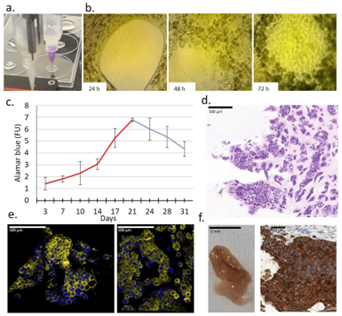
Using alginate-based hydrogel bioink, the researchers bioprinted 3D models of human breast cancer. These models featured a core of breast cancer cells in tissue-like structure. The bioprinted models were cultured long-term to assess cell proliferation in vitro and growth capacity and tumorigenicity in vivo (in SCID mice).
The BioScaffolder 3.2 was employed to create the 3D models, ensuring their accuracy and reproducibility. The results were promising: the 3D bioprinted models demonstrated enhanced cell proliferation and tumorigenic properties compared to 2D cultures. They also provided a more accurate representation of the tumor microenvironment, which is crucial for effective drug testing and metabolic studies.
By using alginate-based hydrogel bioink, the researchers created tissue-mimetic scaffolds that replicate the heterogeneity and drug sensitivity of actual in vivo growing breast tumors. This approach has the potential to reduce the need for animal testing and improve the success rate of clinical trials.
The study concluded that 3D bioprinted breast cancer models offer a more realistic and effective platform for in vitro drug and metabolic targeting. This advancement could lead to better preclinical testing and ultimately improve cancer treatment outcomes.

Taken from Fig 3. Characteristics of a new 3D bioprinted tissue-mimetic model of ZR75.1 human cancer cells. (a) Two dispenser units operated individually for constructing 3D bioprinted tissue-mimetic structures.
This article is based on the following publication:
Dankó, T.; Petővári, G.; Raffay, R.; Sztankovics, D.; Moldvai, D.; Vetlényi, E.; Krencz, I.; Rókusz, A.; Sipos, K.; Visnovitz, T.; et al. Characterisation of 3D Bioprinted Human Breast Cancer Model for In Vitro Drug and Metabolic Targeting. Int. J. Mol. Sci. 2022, 23, 7444. https://doi.org/10.3390/ijms23137444
