Efficient Microalgae Separation Using Dielectrophoresis in a Microfluidic Flow Cell:
Microalgae are a valuable source of poly-unsaturated fatty acids, which are applied in various biotechnological fields such as pharmaceuticals and food supplements. Separating microalgae cells based on their lipid content could be relevant for at-line analytical techniques and strain selection.
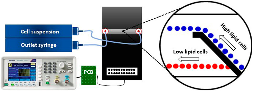
In a study done by researchers from Leibniz-Institut für Innovative Mikroelektronik and Technische Universität Berlin, an electrical approach for the separation of the lipid-producing microalgae Crypthecodinium cohnii using dielectrophoresis (DEP) is explored. The experimental setup involved a microfluidic flow cell with integrated top-bottom electrodes, provided by GeSiM. The single microfluidic channel (500 µm width, 40 µm height) allowed the cell suspension to flow between the electrodes. When an AC electrical signal was applied, the DEP force acted on the cells, deflecting them from their initial path and changing their flow direction.
During 8 days of cultivation, cell growth using optical density was characterized and dry cell weight, glucose concentration and lipid content was characterized via fluorescence microscopy. The size distribution of cells during cultivation was thoroughly investigated, since the DEP force scales with cell volume, but also depends on lipid content via cell electrophysiological constants.
Despite the challenge of deconvoluting separation effects, an efficient DEP-based separation was achieved. The study concluded that dielectrophoresis (DEP) effectively separates cells based on size and electrical properties, correlating with lipid content. While DEP is limited in downstream processes due to low volume turnovers, it can characterize heterogeneous bioproducer populations and aid in strain selection. The non-invasive nature of DEP allows for viable cell separation, making it suitable for handling small, valuable cell quantities and expected to find widespread application in bioprocess engineering.
GeSiM is excited to collaborate on such a wide range of applications, leveraging our expertise to advance biotechnological research and development.

Taken from Fig 6. Schematic of the microfluidic setup used for C. cohnii separation by DEP. The setup consists of a microfluidic flow cell (black platform), two syringe pumps (inlet and outlet), a PCB and a frequency generator. The tubing between the syringe pumps and the microfluidic flow cell is symbolized by light blue-coloured lines and the cables for delivering the DEP signal are indicated by black lines. The magnified view shows the cells’ behaviour due to the DEP field, in which high lipid cells (blue colour) are being repelled at the electrodes edge while low lipid cells (red colour) are passing through the top-bottom electrode structure.
This article is based on the following publication:
Birkholz M, Malti DE, Hartmann S and Neubauer P. Separation of Heterotrophic Microalgae Crypthecodinium cohnii by Dielectrophoresis. Front. Bioeng. Biotechnol. 2022, 10. https://doi.org/10.3389/fbioe.2022.855035
