One Step towards Artifical Liver Models:
The sophisticated tools of the GeSiM bioprinters enable high-level tissue engineering. Here we present a recent paper published by the Centre for Translational Bone, Joint and Soft Tissue Research at the Faculty of Medicine of TU Dresden.
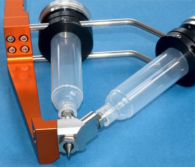
The liver is a pretty complex organ, consisting of different cell types with specific functions, arranged at particular patterns like small “lobules”. Bioengineering of 3D liver constructs is therefore challenging but offering great potential for drug screening/ toxicity testing as well as for disease models. One part of this study was the development of a co-culture system with the Core/Shell extruder on a GeSiM BS3.1 bioprinter. The Core/Shell tool ensures high contact areas between the two different phases.
![Core/Shell structure concept Graphical representation of the hepatocyte-fibroblast co-culture concept, showing hepatocytes encapsulated in the shell are coaxially printed with NIH 3T3 fibroblasts in the core, both co-printed as single cells in their respective bioink (left). An influence of the core composition (with or without fibroblasts and matrix components) on hepatocyte phenotype is hypothesized as they might grow into clusters within their shell compartment and fibroblasts spread to form networks over the cultivation period (right). Images created using Biorender.com software. [1]](https://gesim-bioinstruments-microfluidics.com/wp-content/uploads/2021/03/Scaffold-structure-concept.jpg)
Graphical representation of the hepatocyte-fibroblast co-culture concept, showing hepatocytes encapsulated in the shell, coaxially printed with NIH 3T3 fibroblasts in the core, both co-printed as single cells in their respective bioink (left). An influence of the core composition (with or without fibroblasts and matrix components) on hepatocyte phenotype is hypothesized as they might grow into clusters within their shell compartment and fibroblasts spread to form networks over the cultivation period (right). Images created using Biorender.com software. [1]
It could be shown that the hepatocytes proliferated in all conditions to form multicellular clusters. However, the cluster size was specifically enhanced for co-culture experiments with fibroblasts, simultaneously co-printed in a core/shell fashion.
This proof-of-concept study considered different ink compositions (Alg/MC with and without bioactive components) as well as co-cultures vs. monocultures.
For more information, please download the full publication.
![Metabolic activity after seven days Metabolic activity at day 7 of cultivation (via MTT stain; violet) of HepG2 and NIH 3T3 cells embedded in core–shell printed hydrogel scaffolds; the core–shell interface is visualized via white dashed lines in C. Upper tile shows HepG2 encapsulated in the shell compartment consisting of algMC + Matrigel whereas the core remained cell free. Lower tile shows NIH 3T3 cells encapsulated in the core compartment consisting of algMC whereas the shell remained cell-free. (A,B) Scale bars 2 mm. (C) Scale bars 500 µm. [1]](https://gesim-bioinstruments-microfluidics.com/wp-content/uploads/2021/03/Metabolic-activity-seven-days.jpg)
Metabolic activity at day 7 of cultivation (via MTT stain; violet) of HepG2 and NIH 3T3 cells embedded in core–shell printed hydrogel scaffolds; the core–shell interface is visualized via white dashed lines in C.
Upper tile shows HepG2 encapsulated in the shell compartment consisting of Alg/MC + Matrigel whereas the core remained cell free. Lower tile shows NIH 3T3 cells encapsulated in the core compartment consisting of Alg/MC whereas the shell remained cell-free. (A,B) Scale bars 2 mm. (C) Scale bars 500 µm. [1]
[1] Rania Taymour, David Kilian, Tilman Ahlfeld, Michael Gelinsky & Anja Lode: 3D bioprinting of hepatocytes: core–shell structured co‑cultures with fibroblasts for enhanced functionality, scientific reports, published 4th March 2021


![Cell-free Core/Shell strand Stereo microscopic image of a cell-free core–shell strand, deposited in meandering shape, with clear separation of core (algMC) and shell (algMC + Matrigel) compartments within the strand; scale bar 2 mm. [1]](https://gesim-bioinstruments-microfluidics.com/wp-content/uploads/2021/03/CoreShell_strand_blackwhite.jpg)
![Core/Shell strand with stained core lane Stereo microscopic image of a cell-free, printed scaffold with blue stain in the core for visualizing the continuous core compartment through the coaxial strand; The insert shows a section through the strand segment, cut out from the scaffold scale bar 2 mm. [1]](https://gesim-bioinstruments-microfluidics.com/wp-content/uploads/2021/03/CoreShell_strand_stained.jpg)

