
First in vivo evaluation of 3D-printed FeMn-Akermanite scaffolds for bone repair:
Critical-size bone defects remain difficult to treat because current substitutes rarely combine anatomical fit, mechanical strength, interconnected porosity, and controlled biodegradation. Fe-based biomaterials have shown promise, but traditional manufacturing methods limit their geometric precision and tunability. Additive manufacturing (AM) offers a way to overcome these constraints by enabling porous, multi-material FeMn-Akermanite scaffolds with tailored degradation and bioactivity. Building on earlier in vitro work showing that Fe–Mn–Akermanite (Ak) composites are cytocompatible, osteogenic, MR-friendly, and exhibit improved degradation behavior, this study addresses a major knowledge gap: in vivo studies on AM Fe-based scaffolds are scarce, and in vitro and in vivo results for biodegradable metals are widely acknowledged to correlate poorly. The authors therefore conducted the first in vivo evaluation of this multi-material, extrusion-printed FeMn-Ak composite to assess biodegradation, osseointegration, mechanical integrity, and biosafety in a murine semi-orthotopic critical-size bone defect model.
Two composite formulations (Fe with 35 wt% Mn and either 20 or 30 vol% Ak) were prepared as printable inks and fabricated into porous cylindrical scaffolds using a GeSiM BioScaffolder 3.2. Measured strut and pore dimensions stayed close to the design values (about 3–6% deviation), with 61–63% porosity. After debinding and sintering, scaffolds were characterized by SEM/EDS and μCT. In vitro immersion tests in revised SBF assessed ion release and degradation behaviour. For in vivo evaluation, scaffolds were placed into critical-size defects in cylindrical bovine trabecular bone explants, which were then implanted subcutaneously in nude mice for 16 weeks (four mice, four constructs each). Retrieved constructs underwent μCT, histology, SEM/EDS, and mechanical push-out testing, while major organs were examined for systemic toxicity.
Both scaffold types supported new bone formation and showed excellent osseointegration, with bone growing next to the scaffolds and beginning to infiltrate their pores. In vivo biodegradation was much slower than in vitro (6-8% volume loss after 16 weeks), which the authors believe is due to dense degradation-product layers limiting oxygen transport. Mechanical behaviour differed between the two formulations. FeMn-20Ak scaffolds largely retained their mechanical integrity, with properties in the range of human trabecular bone, while FeMn-30Ak scaffolds, which have a higher ceramic content, became brittle and did not stay intact during testing. No major organ pathology was observed, and only mild iron and manganese accumulation was seen in the liver, which the authors attribute to the high implant-to-body-weight ratio in mice.
Taken together, these preliminary results suggest that extrusion-based AM FeMn-Ak scaffolds can biodegrade slowly, integrate with bone, and, in the FeMn-20Ak formulation, retain mechanical integrity in vivo, marking a step toward their use as biodegradable bone substitutes. FeMn-20Ak emerged as the most promising candidate for its balance of mechanical resilience and osseointegration. The study has clear limitations: four immunodeficient mice, three specimens per group for push-out and histology, and a subcutaneous environment without mechanical loading. The authors therefore call for larger studies in load-bearing orthotopic models in immune-competent animals, with more detailed histopathology, before clinical potential can be assessed. They consider FeMn-20Ak ready for the next stage of preclinical evaluation.
In September 2026, biomaterials researchers gathered at ESB in Antwerp and the DGBM annual meeting in Aachen to share results and move the field forward. As promising materials advance toward preclinical testing, studies like this one offer a useful example of the questions that arise along the way. In vitro, these scaffolds degraded at rates in the range considered suitable for bone, while in vivo degradation was slower and the two compositions differed in mechanical behavior. It is a reminder that degradation rate, mechanical support, and tissue response are best understood together, and that early preclinical results help guide the next steps. We look forward to seeing the community build on these results.
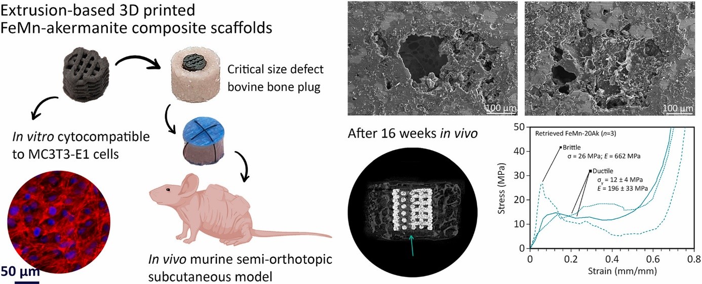
Image taken from Graphical Abstract
This article is based on the following publication: Putra, N.E., Xu, J., Leeflang, M.A. et al. Additively manufactured biodegradable porous FeMn‑akermanite scaffolds for critical‑size bone defects: the first in vivo evaluation. Materials Today Bio 34, 102123 (2025). https://doi.org/10.1016/j.mtbio.2025.102123